
Unravelling the Tangle of Genetic Testing – Part II
by Christian A Scerri MD PhD(Molecular Genetics)
Clinical and Molecular Geneticist
Clinical and Molecular Genetics Clinic
Medical School, St Luke’s Hospital
The Typical Human Gene
Apart from the DNA sequences that can be translated into the corresponding protein, the gene contains or is related to other sequences that control its function and expression. Just upstream of the start codon there is a region that is called the promoter region. This area controls (promotes) the expression of the corresponding gene. Other sequences, this time downstream from the last codon, can also exert control on the gene transcription (enhancing mRNA production). Other important areas are the 5’ and 3’ untranslated regions, the former playing a major role in the control of mRNA translation whilst the latter has an important role in mRNA stability. These regions interact with proteins that are produced by other genes to control the temporal expression of the gene. Thus, the expression of a particular gene is under the influence of other genes within the genome. The rest of the gene is then divided into translated parts (exons) with intervening, non translated parts (introns). Prior to translation, the introns have to be removed (spliced) so that the final mRNA is translated into the protein of interest. Thus, specific sequences that are recognised by the splicing enzyme are present at intron/exon junctions. Another aspect of human genes is that the size can vary from small genes, with a small number of exons (less that 2000 bases, less than 4 exons) to large genes with a large number of exons (more that 1,000,000 bases and more than 30 exons).
What constitutes a Genetic disorder?
Though both environmental and genetic factors play a role in the development of any disease, the definition of a genetic disorder is that of a disease caused by abnormal differences in an individual’s genome. Genetic disorders can be broadly classified in 4 groups:
(1) Single-gene (Mendelian or monogenic) – Single gene disorders are due to mutations in the DNA sequence of one gene. This mutation would result in either a low quantity of the related protein or the production of an abnormal protein that would result in a lack of function of the particular protein. There are over 6,000 identified single-gene disorders with an overall incidence of about 1 out of every 200 births. Some examples are cystic fibrosis, thalassaemia, Marfan syndrome, Huntington’s disease, Duchenne Muscular Dystrophy and hereditary hemochromatosis.
Single gene disorders are inherited in a recognizable pattern – autosomal dominant, autosomal recessive and X-linked. In autosomal dominant conditions, the inheritance of a single mutant allele from either parent would result in the appearance of the disease. A typical example is Huntington’s Disease. In autosomal recessive conditions, the condition would only manifest itself if two mutant alleles are inherited, one from each parent (thus these would be healthy carriers). A typical example of this condition would be cystic fibrosis and thalassaemia. In X-linked disorders, the gene of interest resides within the X-chromosomes. Thus the disease would usually manifest itself in the male progeny of carrier females. Duchene and Becker’s muscular dystrophy are typical examples of these types of genetic disorders.
(2) Multifactorial (complex or polygenic) – This type of genetic condition is by far the most common type as this constitutes most if not all of the chronic and malignant disorders (including the susceptibility of infectious disorders). In this class of disorders, a number of environmental factors and mutations within multiple genes interact together to give rise to the phenotypical picture. Its more complicated nature makes it much more difficult to identify it as a genetic disorder especially when data on the extended family is not available. An example of this condition is Coeliac disease where a clear environment factor (gliadin fraction of the gluten molecule) and genes present on chromosomes 6 (HLA complex), 5 and 19 and the CD28/CTLA4/ICOS gene cluster on chromosome 2 and other as yet to be identified environmental and genetic loci, interact in a still poorly understood way to give rise to the condition. Other examples include breast cancer susceptibility where genes on chromosomes 6, 11, 13, 14, 15, 17 and 22 have been implicated, heart disease, high blood pressure, Alzheimer’s disease, arthritis, diabetes, cancer and obesity.
(3) Chromosomal – In this group of disorders, one finds that large sections of individual chromosomes are missing, duplicated or abnormally rejoined (translocations) and thus result in congenital disease. Most of the major chromosomal abnormalities can be detected by microscopic examination. In most cases the disorder presents at birth in the form of a syndrome. Trisomy 21 (or Down syndrome) is a common disorder that occurs when a person has three copies of chromosome 21.
(4) Mitochondrial – Mitochondria are vestiges of an archeobacterium which were progressively inserted in the cell during evolution. This organelle contains its own, distinct DNA (circular in form) that encodes 37 genes. Mutations within the mitochondrial DNA have been implicated in a number of disorders including cardiomyopathy, diabetes, Friedrich’s Ataxia and optic neuropathy.
Though the knowledge and technology have reached a stage that enable molecular geneticists to study polygenic disorders in an attempt to isolate the causative genes and thus produce clinically applicable tests, up to this date, this is still in its infancy and molecular geneticists are normally faced with the rarer, but easier to define, single gene disorders. Even in these conditions, identifying the causative mutations followed by counselling is fraught with interpretative problems.
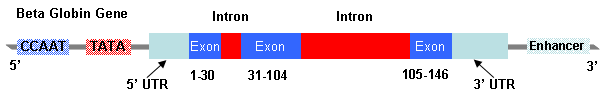
Figure 1. Typical Human Gene (The beta globin gene on chromosome 11)
Bibliography
Ashtar A. Molecular Pathology of Infantile GM1-ganglioasidosis. Malta: University of Malta, 1998.
Avery OT, Macleod CM, McCarty M. Studies on the Chemical Nature of the Substance Inducing Transformation of Pneumococcal Types. J Exp Med 1944; 79:137-58. 1944.
Bezzina-Wettinger S, Balim Z, Felice AE. Allele Frequencies of selected polymorphisms related to thrombosis in the Maltese Population. Blood 2000; 96 part 2: 94b.
Farrugia R, Scerri CA, Montalto SA, Parascandolo R, Neville BG, Felice AE. Molecular genetics of tetrahydrobiopterin (BH4) deficiency in the Maltese population. Mol Genet Metab 2007; 90(3):277-83.
Franklin RE, Gosling RG. Evidence for 2-chain helix in crystalline structure of sodium deoxyribonucleate. Nature 1953;172(4369):156-7.
Galdies R, Pullicino E, Cassar W, Bezzina-Wettinger S, Borg J, Felice AE. A first study on the frequency and phenotypic effects of HFE gene mutations in the Maltese population. Malta Medical Journal 2006; 18[Supplement].
Kellogg DA, Doctor BP, Loebel JE, Nirenberg MW. RNA codons and protein synthesis. IX. Synonym codon recognition by multiple species of valine-, alanine-, and methionine-sRNA. Proc Natl Acad Sci USA 1966; 55(4):912-9.
Mullis K, Faloona F, Scharf S, Saiki R, Horn G, Erlich H. Specific enzymatic amplification of DNA in vitro: the polymerase chain reaction. Cold Spring Harb Symp Quant Biol 1986;51 Pt 1:263-73.
Scerri CA. Clinical and Molecular Pathology of the beta+ IVSI-6C Thalassaemia in Malta. Malta: University of Malta, 1998.
Vella J. The Detection of the DNA Mutations that Cause Gangliosidosis. Malta: University of Malta, 2000.
Watson JD, Crick FH. The structure of DNA. Cold Spring Harb Symp Quant Biol 1953; 18:123-31.
Wilkins MH, Seeds WE, Stokes AR, Wilson HR. Helical structure of crystalline deoxypentose nucleic acid. Nature 1953;172(4382):759-62.