
Imaging Breast Implant Rupture – Part II
Dr Pierre Vassallo
Ultrasound
Breast ultrasound is superior to mammography for detection of breast implant leaks but less accurate than breast MRI. Given the wide availability and low cost of breast ultrasound compared to breast MRI, it has become a very important tool. Since its negative predictive value for detecting leaks is high,4 breast ultrasound is often used as a first examination before proceeding to MRI for more accurate assessment of prosthesis integrity.
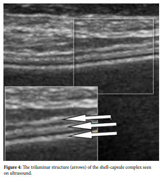
A single lumen silicone implant appears anechoic with no internal features on ultrasound. Implants fold themselves within the surgical pocket created by the plastic surgeon; these folds should not be mistaken for implant leaks. With time, a fibrous capsule forms around the implant; this capsule and the implant shell form a capsule-shell complex that appears as three parallel echogenic lines on ultrasound (Fig 4).
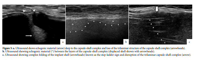
Intracapsular leaks may appear on ultrasound as echogenic material deep to the capsule or as an interruption of the capsuleshell complex (Fig 5a). They may also present as echogenic material between the layers of the capsule-shell complex (Fig 5b). An intracapsular tear may also result in complex folding of the implant shell known as the step-ladder sign (Fig 5c). It is important not to confuse normal implant folds with an intracapsular leak.
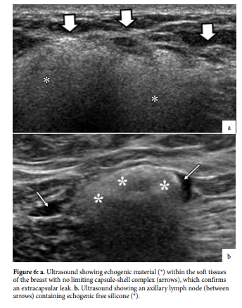
An extracapsular leak presents as echogenic material (silicone) within the soft tissues of the breast with no delimiting trilaminar complex (Fig 6a). Free silicone may also be present in the axillary lymph nodes (Fig 6b).
Breast MRI
MRI is the most accurate imaging modality to assess breast implant integrity. In the US, the food and drug administration recommends a breast MRI three years after implant surgery and bi-yearly thereafter to monitor implant integrity. However, this is not universally accepted since there is no clear evidence that it will influence patient morbidity.
Careful questioning of patients prior to breast MRI is required; saline-filled implants do not require MRI evaluation, while the presence of tissue expanders (implants that can be filled by external injection of saline) are a contraindication to MRI, because they contain magnets at the injection port. Only siliconefilled implants should undergo MRI examination.
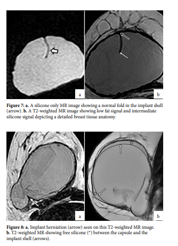
The augmented breast contains fat, water and silicone, and MRI can analyse each of these components separately clearly mapping each one within the breast. MRI sequences that null out fat and water clearly depict extracapsular silicone (Fig 7a), while sequences that null out silicone can distinguish a silicone leak from a fluid collection (Fig 7b).
MRI allows accurate assessment of the posterior margin of the implant, which is difficult to see on ultrasound. Implant herniations through the capsule are best seen on MRI and although they do not constitute a leak, they will result in contour deformity (Fig 8a). The presence of free silicone between the implant shell and the capsule can readily confirm an intracapsular rupture (Fig 8b). On the other hand, the classical “linguine” sign, which correlates with the complex folds of the collapsed implant shell, may also occur with intracapsular rupture (Fig 8c).
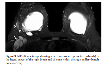
Extracapsular tears and the presence of free silicone in the axillary tissues and lymph nodes can be readily evaluated with silicone selective MR imaging (Fig 9). Implant assessment MR protocols must be clearly distinguished from breast cancer screening protocols. The latter require injection of intravenous contrast agent. However, both protocols can be combined if required, delivering the best analysis of implant integrity and the most accurate screening method for breast cancer.
Extracapsular silicone leaks may sometimes mimic breast cancer on mammography and ultrasound; breast MRI can distinguish the two entities and therefore is a valuable tool when assessing patients with a high-risk for breast cancer who have had breast augmentation.
The new generation of breast implants are composed of semisolid silicone gel (cohesive or “gummy bear” implants). These designs are aimed at reducing the risk of free silicone migration into soft tissue. These implants have been noted to fracture rather than leak; these fractures are best evaluated with breast MRI.
Conclusion
Breast imaging is one of the most commonly performed diagnostic imaging studies. Although breast imaging is mainly aimed at detecting early breast cancer, an increasing number of women who attend breast cancer screening have had breast augmentation procedures. It is important to recognise the radiological findings related to breast implant leaks as they may mimic breast cancer. Breast implant imaging is also important when planning management of implant leaks.
References cont.
- Venta LA, Salomon CG, Flisak ME, et al. Sonographic signs of breast implant rupture. AJR Am J Roentgenol 1996;166(6):1413–1419.