
Bacterial Conjunctivitis
Melvin J. Gouder
Introduction
Bacterial conjunctivitis is a very common ophthalmic condition that can affect anyone from day one of life to old age1. It is one of the commonest reasons for self-referrals of patients visiting eye specialists. It is defined as an inflammation of the conjunctiva by bacteria where the palpebral, bulbar and forniceal parts of the conjunctiva become hyperaemic. The infection can be acute, hyperacute or chronic. Contrary to popular belief it can be self-limiting but it is frequently treated with broad-spectrum antibiotics most commonly in drop form2. Very rarely it can progress to complications such as keratitis (corneal infection) or pre-septal cellulitis (skin infection of the lids)3.
Causation
There is a whole host of pathogenic bacteria that can cause this kind of benign ocular pathology4. The spectrum of bacteria includes Neisseria, Staphylococcus Haemphilus, Moraxella and Chlamydia species, as well as thehighly virulent form of Streptococcus pneumoniae. The acute form is commonly caused by Staphylococcus aureus, Streptococcus pneumoniae or one of the Haemophilus species. Moraxella and Chlamydia can cause a chronic form. Clinically, Chlamydia can cause a non-purulent form of conjunctivitis with symptoms more synonymous of a viral infection like adenoviral conjunctivitis.
Risks
The most common risk factor to acquire all forms of bacterial conjunctivitis is coming in contact with an infected individual5. However other risks include poor drainage of tears such as in nasolacrimal duct obstruction (NLDO), lid malposition such as entropion or ectropion (also interfering with lacrimal drainage), severe tear deficiency as in Sjogren syndrome and the associated autoimmune diseases. These conditions hinder the natural resistance mechanisms of the eyes. Patients on immunosuppressive agents such as steroids are at greater risk, as are older fragile patients. Good hand hygiene and limiting direct contact with infected individuals reduce the risks.
Symptomatology and signs of bacterial conjunctivitis
The differential diagnosis of bacterial conjunctivitis includes its viral form, the allergic variant and less commonly, conjunctivitis secondary to chemical exposure. All the signs and symptoms can be similar but in bacterial conjunctivitis there are some specific features. A purulent discharge that can have mucoid characteristics, irritation, diffuse conjunctival hyperaemia and bulbar conjunctival injection are all features that occur first in one eye but then commonly spread to the fellow eye.
Diagnosis
The diagnosis is most commonly clinical1. The patients’ description that they might have come in contact with an infected individual also helps diagnosis. Full ophthalmic examination is necessary, including slit-lamp biomicroscopy to exclude intraocular pathology. If there is an associated keratitis one might observe a secondary reaction in the anterior chamber which may warrant treatment. Additional diagnostic tests are usually reserved for recurrent and/or chronic cases which are unresponsive to the initial medication. These include cultures, stains, smears and immunoassays. Typically in a chronic conjunctivitis one might need to specifically screen for viral causes (Herpes) and Chlamydia apart from cultures for the other forms of stubborn bacteria.
Treatment
The gold-standard treatment is with a broad-spectrum antibiotic in drop or ointment form for 5-7 days and is commonly effective. As mentioned above, it can also be self-limiting but treatment may reduce the symptoms and duration of the disease. Recurrence rates are lower in treated cases.
Table 1. Commonly used antibiotics in bacterial conjunctivitis in Malta
| Antibiotic group | Example | Mode of Action |
| Aminoglycosides |
Gentamicin Tobramycin |
Protein synthesis inhibition |
| Chloramphenicol | Chloramphenicol | Protein synthesis inhibition |
| Fluoroquinolones |
Ciprofloxacin Moxifloxacin |
Interferes with DNA synthesis |
Sometimes aminoglycosides are not considered to be broad-spectrum antibiotics due to their poor activity against Streptococci6. Chloramphenicol resistance has become very common so it is not popular amongst ophthalmic surgeons5. On the other hand, erythromycin ointment is not commonly prescribed but is usually reserved for Chlamydial infections. It was traditionally a hospital item.
Fluoroquinolones are in their 4th generation (moxifloxacin) and can also be used in patients with infection related to contact lens misuse such as keratitis.
Conclusion
Though benign, bacterial conjunctivitis should never be treated lightly especially in older patients who are to undergo cataract surgery. Any suspicion of infection in these patients should be initially treated with topical antibiotics. One should wait clearance from sensitivity tests to ensure that the eye is free from pathogenic bacteria that can potentially cause acute post-operative bacterial endophthalmitis. There are ophthalmic surgeons who prefer to treat high-risk patients (diabetics and the immunocompromised) with a pre-operative course of 4 days with a 4th generation fluorinated quinolone like moxifloxacin.
References
- American Academy of Ophthalmology Cornea/External Disease Panel, Preferred Practice Patterns Guidelines. Conjunctivitis – Limited Revision. San Francisco: American Academy of Ophthalmology; 2011. Available from: guideline.gov/content.aspx?id=36093
2. Azari AA, Barney NP. Conjunctivitis: a systematic review of diagnosis and treatment. JAMA 2013; 310(16):1721-9. doi: 10.1001/jama.2013.280318.
- Conjunctivitis – information prescription. Complications of conjunctivitis UK NHS. Available from: nhs.uk/Conditions/Conjunctivitis-infective/Pages/Complications.aspx
4. Watkinson S. Assessment and management of patients with acute red eye. Nurs Older People 2013;25(5):27-34; quiz 35. PubMed PMID: 23914708.
5. Mishori R, McClaskey EL, WinklerPrins VJ. Chlamydia trachomatis infections: screening, diagnosis, and management. Am Fam Physician 2012; 86(12):1127-32.
- Karpecki P, Paterno MR, Comstock TL. Limitations of current antibiotic treatment of bacterial conjunctivitis. Optom Vis Sci 2010; 87(11):908-19.
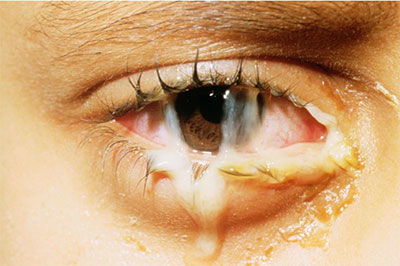
Figure 1. Bacterial conjunctivitis showing prominent mucopurulent discharge, inflamed bulbar conjunctiva and lid swelling